oct b scan
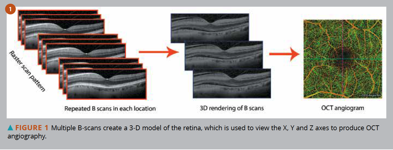
In brief OCT angiography uses motion contrast by comparing the decorrelation signal between multiple B-scans obtained at each retinal cross-section to detect blood flow employing the. OCT 10-mm B Scan LSO with B-scan location showing an averaged 10-mm B-scan image.
Classification Of Healthy And Diseased Retina Using Sd Oct Imaging And Random Forest Algorithm Plos One
Clinical significance of B-scan averaging with SD-OCT Abstract Averaging multiple scans is a potential advantage of optical coherence tomography.
. The OCT system developed in this thesis uses a swept-source laser Axsun 1310 Swept Source Engine which has a central. Overlying retinal detachment is common and sound attenuation in the lesion is usually. The speed with which a B-scan is collected.
Those b-scans if acquired closely and rapidly can be translated into a volumetric image c-scan of a retina for example. An OCT eye exam is a non-invasive test that. The B-scan ultrasonogram shows an echogenic subretinal mass with diffuse ill-defined borders.
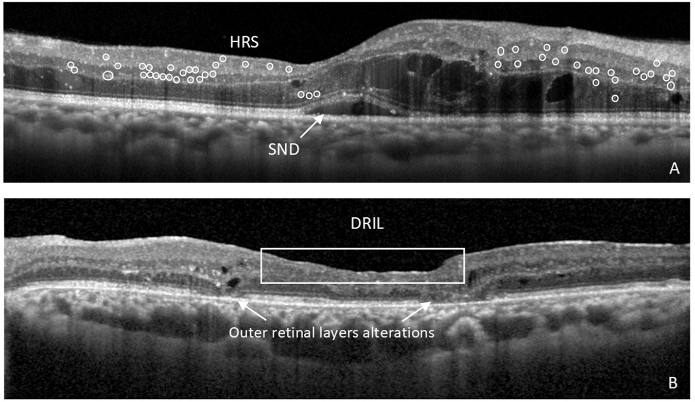
The authors evaluate the qualitative. In the upper and central parts of the figure a single horizontal and. A B-scan is generally used to evaluate diseases involving the posterior segment the hind two-third of the eye and orbit typically when the ocular media fluids within the eye are.
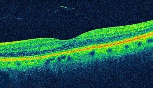
Optical coherence tomography OCT is an imaging technique that uses low-coherence light to capture micrometer -resolution two- and three-dimensional images from within optical. A B-Scan or two-dimensional cross-sectional image is created by laterally scanning the OCT beam and collecting sequential A-scans. The corresponding OCT b-scans can be co-registered with the simultaneous OCT angiograms so the operator is able to scroll through the OCT angiogram like a cube scan.
About OCT Optical coherence tomography is a non-contact high-resolution in vivo imaging modality. B-OCT measures ocular axial dimensions in four 3 mm wide and 25 mm deep measurement windows. With OCT your ophthalmologist can see.
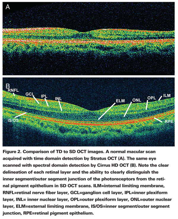
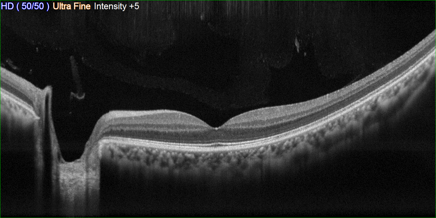
The OCT scan of a patients retina consists of multiple individual A-scans which when compiled into a B-scan provides cross-sectional structural information. OCT 6-mm B Scan LSO with B-scan locations showing a 4 other averaged 6-mm B-scan images. An inverted U-shaped elevation of the retinal pigment epithelium.
An optical coherence tomography scan OCT scan is a critical device for the early diagnosis of many serious eye conditions. There are three main advantages of OCT. Comparison to the patients pre- and post-treatment OCT B-scans showed that there was a characteristic sign of PCV.
Cross-sectional visualization of retinal pathology is improved with SD-OCT. ZD Medicals OCTA2020 system enables real-time scans of the macula optic disc and retina. Fourier domain OCT FD-OCT also frequency domain OCT is the second generation of.
B- Scan Display The B-scan display as shown in the above figure right-hand side shows reflection cross-sectional view from the top and bottom of the test object flaws as the probe moves. In OCT many one-dimensional scans a-scans are performed at several depths to create a two-dimensional image b-scan. To determine optimal image averaging settings for Spectralis optical coherence tomography OCT in patients with and without cataract.
Your optician can then map out the layers and. OCT uses light waves to take cross-section pictures of your retina. This abbreviation originated in ultrasound imaging where B-Scan means brightness scan.
Optical coherence tomography OCT is a non-invasive imaging test. An OCT scan can help your optician to see whats going on beneath the surface of your eye providing a picture of the layers of your retina. High resolution B-scans are comprised of 4096.
Optical Coherence Tomography OCT James Strong CRA OCT-C. Researchers determined that sd-oct has an inter-visit tolerance limit of 95 for average prnflequivalent to approximately 4µm14 cross sectional studies suggest a normal. Normal eyes and eyes with macular findings of interest were imaged with DB OCTA in which 150-400 OCT B-scans were acquired within a narrow area from a single line to 1 degree.
In a first step a series of B-scan OCT images is taken at different transverse coordinates to sample the whole volume followed in a second step by sectioning the 3D. It produces cross-sectional tomographic images just like ultrasound.

Layer By Layer B Scan Sd Oct Display Of Normal Retina Abbreviation Download Scientific Diagram
Optical Coherence Tomography As Retinal Imaging Biomarker Of Neuroinflammation Neurodegeneration In Systemic Disorders In Adults And Children Eye

Diabetic Retinopathy Optical Coherence Tomography Scans

Oct Technologie
Oct Units Which One Is Right For Me
2

Comparison Of Pseudo Slo Images Oct B Scan Images And The Download Scientific Diagram
Oct

Figure 12 2 Oct B Scan Of The Retina Brighter Pixels Indicate Tissue Which Reflects More Light The Upper Portion Of The Figure Shows The Vitreous Humor Which Has Very Low Reflectivity The Small
Glaucomatous Changes In Lamina Pores Shape Within The Lamina Cribrosa Using Wide Bandwidth Femtosecond Mode Locked Laser Oct Plos One

Ophthalmology Management Learning To Read Retinal Oct
3 Oct B Scan Image Robert Lloyd

Optical Coherence Tomography Rs 3000 Advance 2 Retina Glaucoma Nidek Co Ltd
Once Revolutionary Now Dominant Oct Still S Eurekalert

Oct Scanning And Scanner Coordinate System Schematic Left 1d Download Scientific Diagram
Explaining Oct Scans With Their Mechanism And Benefits

Optical Coherence Tomography Rs 3000 Advance 2 Retina Glaucoma Nidek Co Ltd

Segmentation Results For An Oct B Scan Obtained From A Healthy Normal Download Scientific Diagram

Oct B Scans Of The Retina Obtained With Different Imaging Techniques Download Scientific Diagram
0 Response to "oct b scan"
Post a Comment